MRI
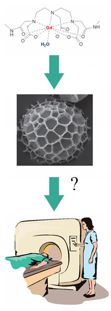
The principle behind this technique is that the exine shells concentrate the contrast agent in a particular part of the body, where it then shows up brightly within the scanner.
 There are three potential benefits from using this technique:
- The exines go to parts of the body not normally reached by contrast agents such as gadolinium
- The contrast agent is released in blood over a period of time so sequential imaging
- A much smaller quantity of contrast agent is required
We have demonstrated that these microcapsules can be filled with a commercial gadolinium(III) MRI contrast agent which was slowly released in blood plasma due to enzymatic digestion of the exine capsule. Magnetic Resonance Microimaging Experiments were carried out at 11.47 T to characterise the release profile over time of the commercial contrast agent (CA) Omniscan. The loading level of the Omniscan was determined by ICP-OES to be 0.80%. The figure below shows four consecutive slices through the agar gel monolith containing Omniscant encapsulated in exine shells. Each frame has dimensions of 3 x 3 mm. An exine capsule that appears in several consecutive slices is highlighted with arrows here.
The plot here shows the release of the contrast agent over 8 hours. A maximum is reached after ca. 4 hours for the sample in human blood plasma and no significant release is observed for the control sample.
More information on this experiment is given in M. Lorch, M. J. Thomasson, A. Diego-Taboada, S. Barrier, S. L. Atkin,G. Mackenzie and S. J. Archibald, Chem. Commun., 2009, DOI: 10.1039/b909551a
This technique can also be applied to non-conventional contrast agents such as food oils. The picture shown below was obtained after drinking milk containing the exine shell loaded with fish oil.

Photograph: Ten minutes after the ingestion of oil filled exine. Stomach mucosa highlighted by exine (arrow) and increased signal in the small vessels of the liver
– shown in the region of interest highlighted by box.